THE MIYA MODEL STORY
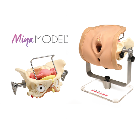
The MIYA Model consists of a pelvic frame and multiple replaceable anatomic cartridges and is designed with a number of specific surgical procedures in mind. The model incorporates a number of features to simulate actual surgical experiences including lifelike skin and life-size organs, realistic cutting and puncturing tensions, palpable surgical landmarks, a pressurized vascular system with bleeding for inadequate technique, and an inflatable bladder that can leak water if damaged.
Mounted on a rotating stand with the top of the pelvis open, the MIYA Model is designed to provide access and visibility, enabling supervising physicians and credentialing committees with topical access and video capabilities, facilitating greater guidance and review. The light weight model offers convenient transportation, set-up, use, and storage. Today, the MIYA Model can be used to practice all major vaginal surgical procedures with the potential to support additional procedures in the future.
The current standard for vaginal surgery instruction is reliant upon both live surgery and the use of training cadavers. These approaches suffer from several handicaps, including: patient risks, liability risks, availability limitations, high costs and high demand on proctoring surgeons. Over time, these limitations have caused a decrease in resident exposure to live training cases. In fact, Dr. Miyazaki conducted research at his home institution, finding the number of hysterectomies performed by residents has drastically decreased. Current residents are performing about a quarter of the number of hysterectomies Dr. Miyazaki performed during his own residency, just two decades ago. Trends in health care and a growing shortage in funding dollars suggest that this alarming dynamic will only continue to get worse.
These issues are not institution specific and have become an ever growing problem in residency programs across the country. Consequently, dramatic declines in surgical experience are generating a growing interest in the objective verification of minimal competency levels as residents graduate from one level to the next, prompting the Accreditation Council for Graduate Medical Education (ACGME) and American Congress of Obstetricians and Gynecologists (ACOG) to develop and implement the Milestone Project. Despite the demands for increasing the quantity and quality of surgical training, as well as growing requirements for competency assessment and verification, neither the status quo of live surgeries and cadavers, nor other alternatives such as existing surgical simulators or anatomical models, are adequate solutions given the cost constrained environment.
The market demanded something different–a training tool that would provide lifelike quality, tactile feel, and meticulous anatomical placement, all while delivering cost efficiency, mobility, a low-risk environment, reproducibility, repetition of procedures and high-fidelity training–every time. After assessing these variables, Dr. Miyazaki determined that if designed appropriately, model-based simulation training could provide an ideal solution. He then put his innovative and entrepreneurial skills to work, and after more than six years of intense research, discovery, product development, testing, and validation, the MIYA Model was born.
Supported Training & Procedures
The Miya Model is the only surgical simulation model that can support such a wide range of surgical training and procedures with the potential to add more in the future.

Basic Gynecological Examination
- Speculum exam
- Cervical visualization and pap smear
- Endometrial biopsy

Palpation of Key Surgical Landmarks
- Pubic tubercles
- Pubic rami
- Obturator fossa
- Arcus Tendineous Fascia Pelvis
- Ischial Spine
- Sacrospinous Ligament

Basic Gynecological Examination
- Total Vaginal Hysterectomy with or without Bilateral Salpingo oophorectomy
- Transoburator sling placement, both outside-in and inside-out approaches
- Retropubic sling placement, both top-down and bottom-up approaches
- Full thickness vaginal wall dissection to the Arcus Tendonius Fascia Pelvis
- Bilateral Sacrospinous Ligament Fixation Dilation and Curettage
- Uterine morcellation
- Dilation and Curettage
- Diagnostic Hysteroscopy
Miya Model Components
The MIYA Model base model and cartridges were meticulously designed with high quality materials and carefully designed for realism, durability, and cost effectiveness.

Frame & Pelvis
- Weighted stand with support beams designed to fit on a desktop
- Support brackets can Rotate 360° to enhance teaching
- Enables pelvis to be placed in Trendelenburg and reverse Trendelenburg positions to simulate real surgical table positioning
- Model easily assembled
- Model to be used anywhere without any special set up
- Compact storage
- Pelvis specially designed and included as part of the base model to fit multiple cartridges for precise anatomic relationships and to to simulate real tissue pathology
- The pelvis and the base are of both made of a hard plastic composite for durability
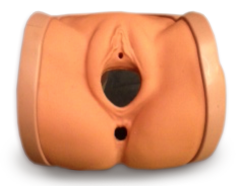
Vulva Assembly
- Realistic appearance and feel. Flesh toned with darker rectum as visual aid
- Includes lower stomach, vulva, urethral opening, vaginal introitus, anus and rectum
- Lower abdominal skin allows for bimanual exam and retropubic sling passage
- Soft, lifelike feel allows for palpation of bony landmarks, including: pubic tubercles, pubic rami, obturator fossa, ischial spine, ischial tuberosity and coccyx
- Sandwiched between inner and outer frame to support outer body shape and stability against pelvis
- Rectum is a hollow tube allowing digital exam
- Vulva skin thicker in some areas to simulate fatty tissue
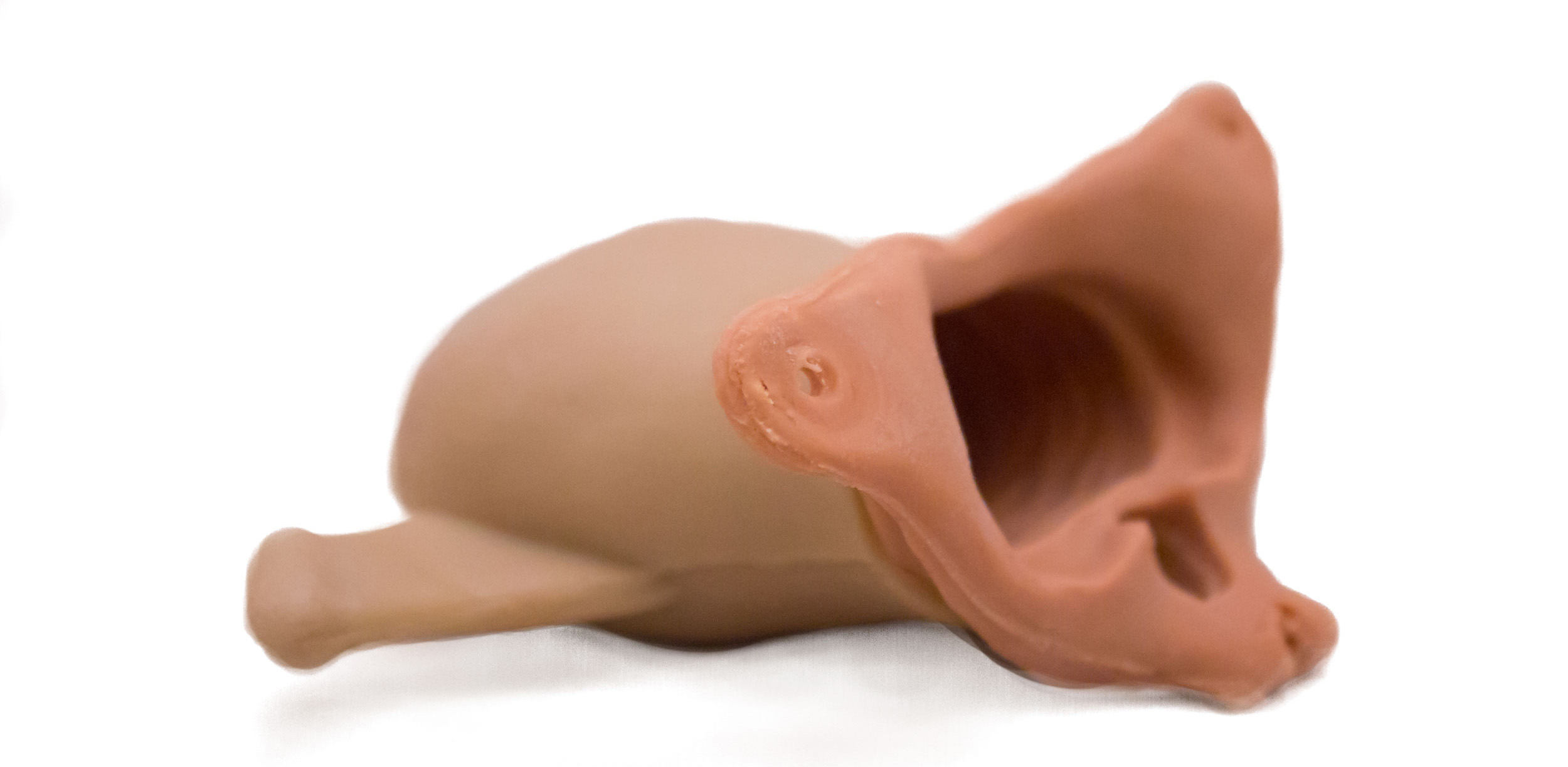
Vagina with Endopelvic Fascia Layer
- Realistic feel and vaginal texture
- Soft vaginal wall allows for palpation of Arcus Tendonius Fascia Pelvis (ATFP) from inferior pubic ramus to the ischial spine
- Special materials design enables the surgeon to have a realistic feel when cutting with scissors or scalpel and suturing the vaginal wall
- Opens from vulva at pubic rami, vagina widens and goes from arcus to arcus bilaterally, almost to spines stopping about 1 cm before spinous process
- Anterior and Posterior cul-de-sacs are palpable
- Endopelvic fascial layer extends to ATFP and is anchored to a slot above the ischial spines of the pelvis and creates fascia plane and enables full thickness vaginal wall dissection to the Arcus Tendonius Fascia Pelvis
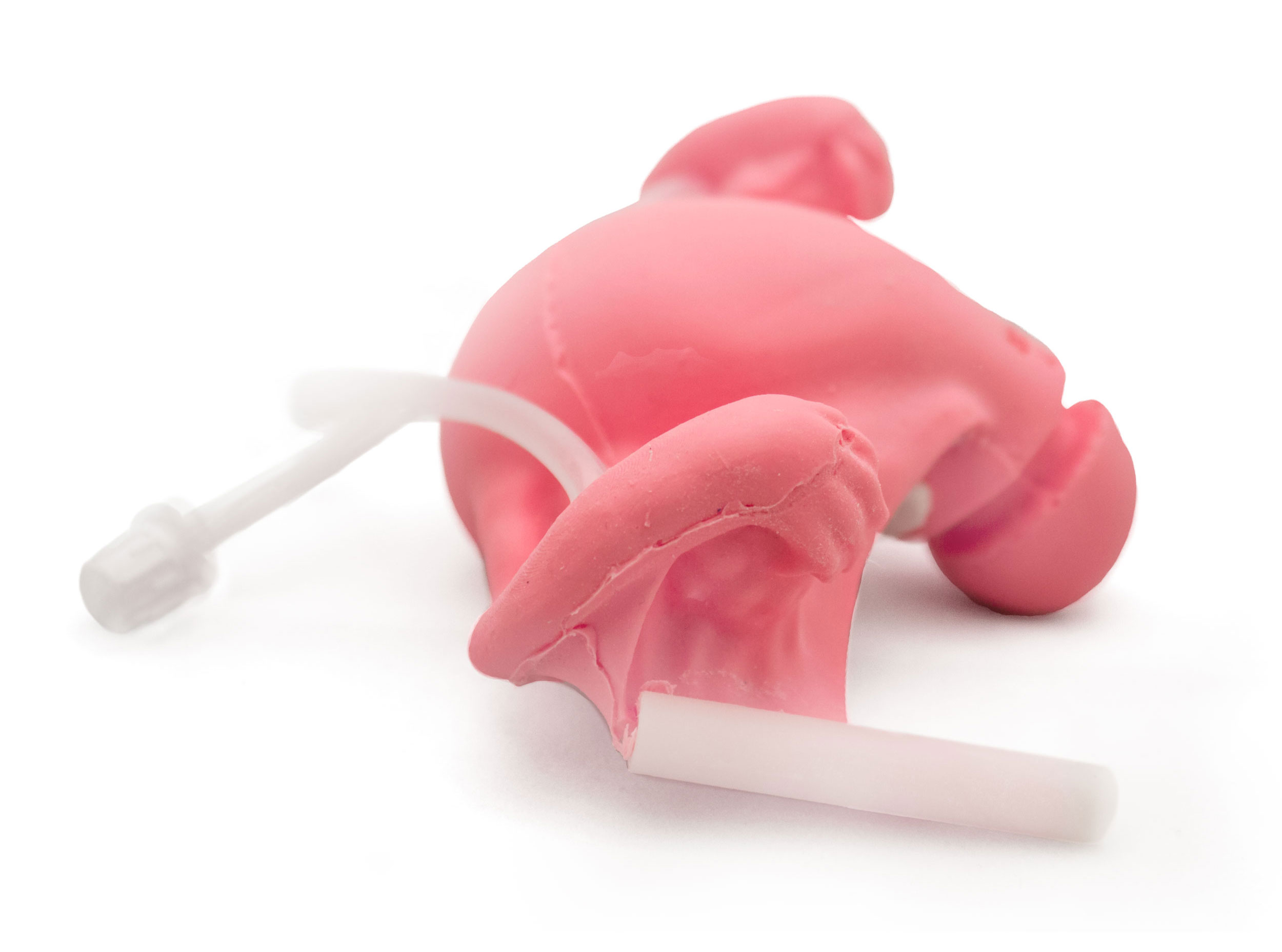
Uterus Tubes & Ovaries
- Two versions of uterus available, one for vaginal hysterectomy surgery training and another for more general usage
- Includes cervix, fallopian tubes and ovaries
- Realistic cervical os connecting to full uterine cavity, accepts IUD
- Broad ligament with connections to pelvic frame via stiff rods for secure attachment
- Easy replacement into sliding lock groove in pelvis
- Vessels run through broad ligament and connect via luer locks to simulate vascular supply; will fit standard IV tubing
- Uterine cavity is normal gynecoid shaped and is currently designed for IUD insertion and removal, endometrial biopsy, dilation and curettage, and diagnostic hysteroscopy
- Vaginal hysterectomy with or without bilateral salpingo oophorectomy can also be performed, the surgeon is able to perform a complete procedure on the model
- The normal bony pelvis and anatomy further enhances the surgical experience with clamp placement technique, suturing and knot tying
- Uterine morcellation is able to be performed
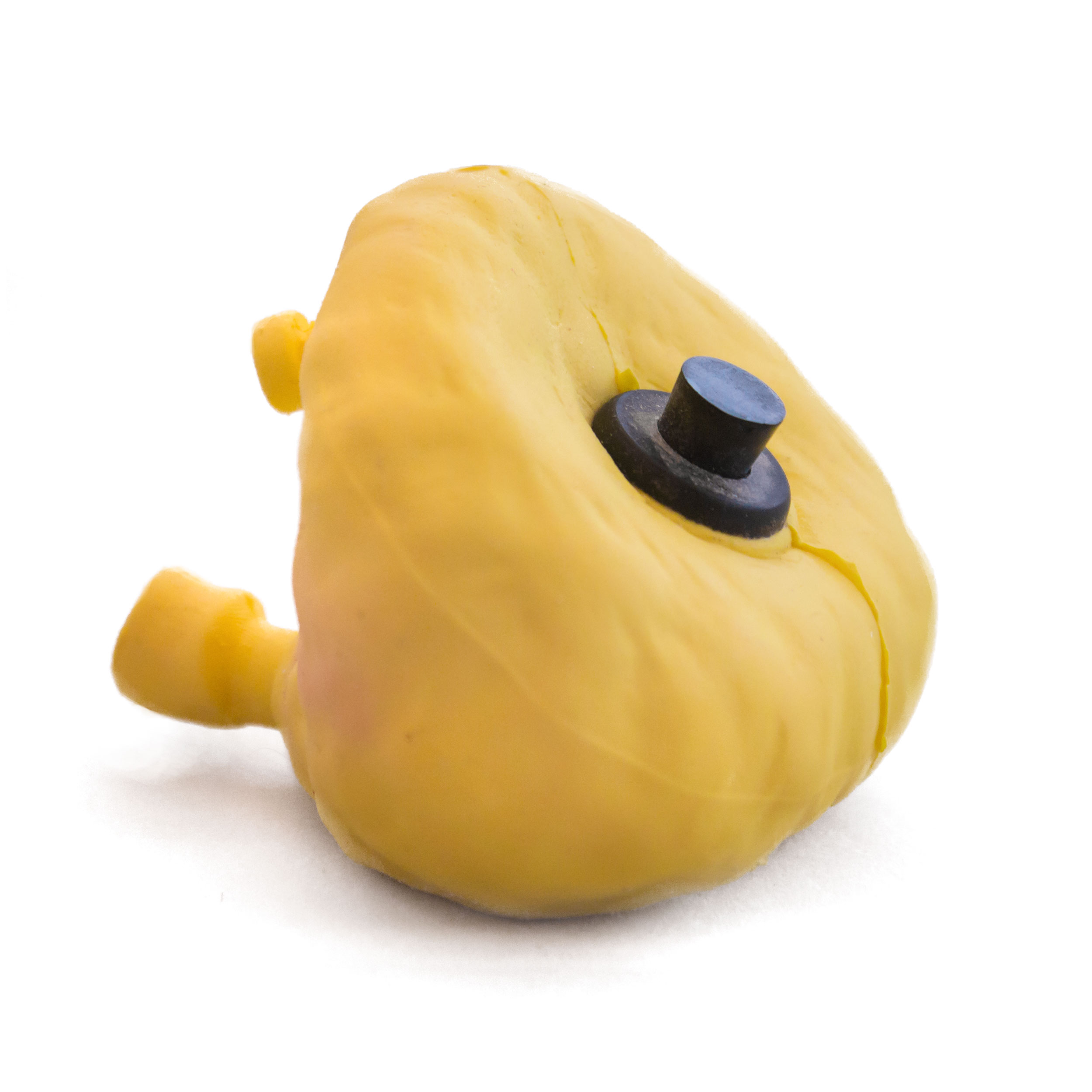
Bladder
- Hollow bladder with opening on top for filling with water, grommet and plug
- Expands to hold 100 cc of fluid, leaks when punctured
- Deflates when empty
- One way valve in urethra simulates sphincter and allows for cauterization, accepts 12-16fr catheter
- Attaches anteriorly to groove in pubic bone and fits snugly between pubic bone and uterus

Sacrospinous Ligament
- Single band with hook connections at each ischial spine and screw on sacral plate
- Simulates ‘pop’ of puncture with trocar passage
- SS ligament has the “characteristic” ridges which are palpable on the vaginal and rectal exam
- The ligament originates from the ischial spine and terminates into the lateral sacrum
- Trocar/needle passage can be from above or below the ligament
- Sacral edge lines up with foramina S4 S5, about 3 cm wide as per cadaver studies, prominent texture can be felt through the vaginal wall.
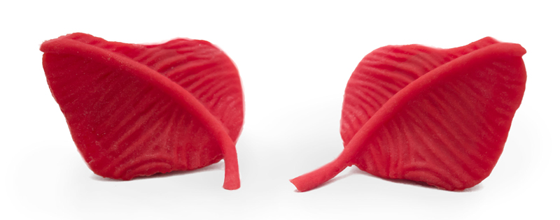
Obturator Complex
- Combined obturator membrane and obturator internus/externus muscles
- Obturator fossa is easily palpated
- 360 degrees of tension during palpation
- Obturator complex is housed with a circular frame which turns and locks into the pelvic sidewall at the obturator foramen
- Special locking mechanism allows for realistic trocar passage feel and “pop” from an inside out or outside in approach
- Includes raised ridge of the ATFP which extends from the inferior pubic rami towards the ischial spine.
- Anchoring devices and suturing can be performed along the length of the Arcus Tendonius Fascia Pelvis
- Simulates “pop” of puncture

Perineum
- Combined obturator membrane and obturator internus/externus muscles
- Obturator fossa is easily palpated
- 360 degrees of tension during palpation
- Obturator complex is housed with a circular frame which turns and locks into the pelvic sidewall at the obturator foramen
- Special locking mechanism allows for realistic trocar passage feel and “pop” from an inside out or outside in approach
- Includes raised ridge of the ATFP which extends from the inferior pubic rami towards the ischial spine.
- Anchoring devices and suturing can be performed along the length of the Arcus Tendonius Fascia Pelvis
- Simulates “pop” of puncture


